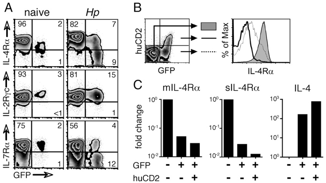
FIGURE 6.
Highly activated Th2 cells lose IL-4Rα expression. A, WT 4get mice were infected with H. polygyrus, and 14 d later, CD4+ cells from mesLNs were analyzed for their expression of IL-4Rα, IL-2Rγc, and IL-7Rα. B, 4get/Kn2 dual-reporter mice were also infected, and on day 14, mesLN CD4+ cells were sorted into three populations as shown (GFP− huCD2−, filled histogram; GFP+ huCD2−, dashed line; GFP+ huCD2+, solid line). IL-4Rα expression on each population was measured by flow cytometry. C, Populations sorted as in B were lysed, their RNA was purified and reverse-transcribed, and their expression of the membrane-associated and soluble alternative splice variants of the IL-4Rα transcript and of IL-4 was assessed by quantitative PCR. Data are shown as a fold change in expression relative to that of GFP− huCD2− cells. In all of the panels, data are representative of at least two independent experiments.

