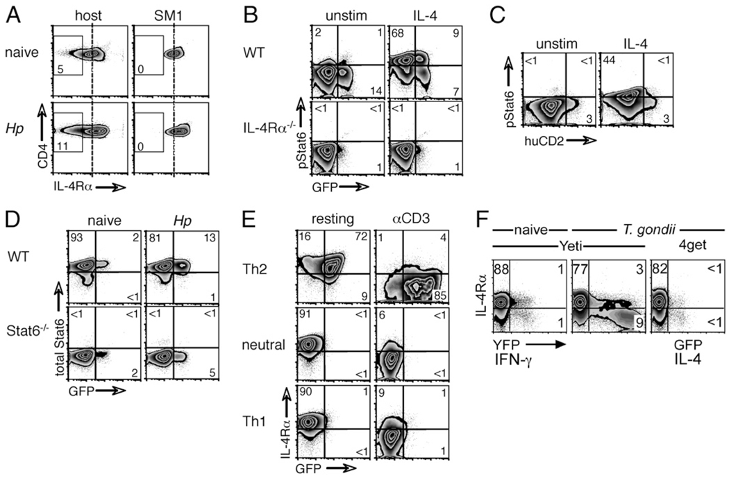
FIGURE 7.
IL-4Rα downregulation is triggered by Ag encounter and terminates IL-4R signaling. A, Mixed BM chimeras were generated by reconstituting WT CD90.2+ C57BL/6 recipients with a 90:10 mix of WT CD90.2+ and SM1 TCR transgenic CD90.1+ BM. Chimeras were infected with H. polygyrus, and 14 d later, CD4+ cells from mesLNs were analyzed for IL-4Rα expression by flow cytometry. Data shown are gated on CD4+ cells, and the numbers indicate the percentage of IL-4Rαlo cells within each population. B, WT and IL-4Rα−/− 4get mice were infected with H. polygyrus, and mesLN cells were isolated on day 14. Cells were first incubated with anti–IL-4 to remove endogenous STAT6 phosphorylation and then either incubated in medium alone or stimulated with IL-4 for 15 min. Resulting STAT6 phosphorylation was assessed by intracellular staining and flow cytometry. C, WT 4get/Kn2 dual-reporter mice were infected with H. polygyrus, and mesLN cells were harvested, stimulated, and stained as in B. D, WT and STAT6−/− 4get mice were infected with H. polygyrus, and mesLN cells were isolated on day 14. Total STAT6 expression was assessed by intracellular staining and flow cytometry. E, Naive CD4+ cells from 4get mice were activated with αCD3 + αCD28 in Th2, neutral, or Th1 conditions. Cells were washed, rested, and on day 7 either cultured in medium alone or restimulated with αCD3. IL-4Rα expression was measured 24 h later by flow cytometry. F, Yeti IFN-γ and 4get IL-4 reporter mice were infected with the Th1-inducing protozoan T. gondii, and 7 d later, CD4+ cells from mesLNs were analyzed for their expression of YFP or GFP and IL-4Rα. Data in all of the panels are representative of at least two independent experiments.

