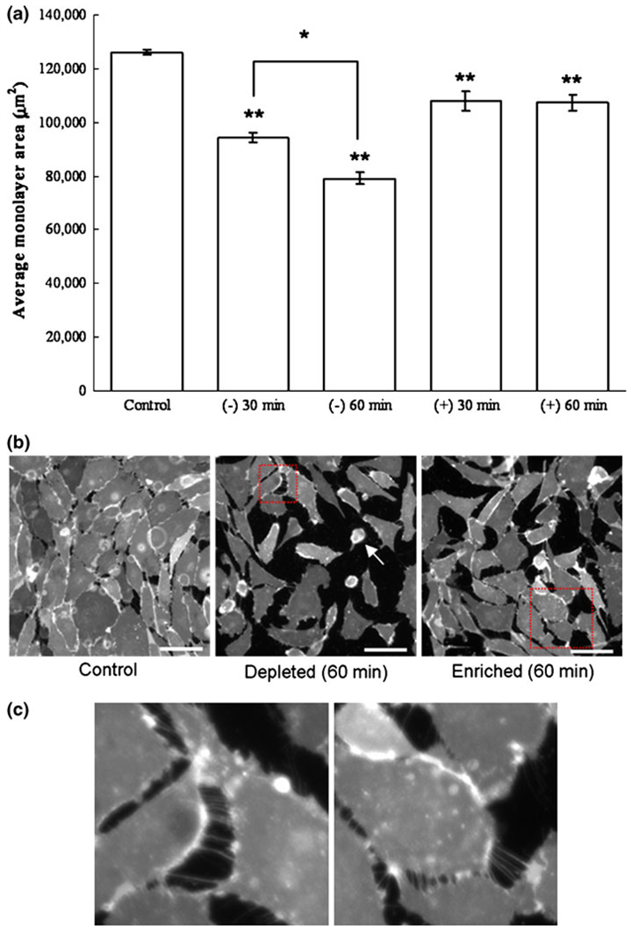
FIGURE 5.
Effect of cholesterol treatment on BAEC confluent monolayer plated on a fibronectin-coated glass for control, 30 min, and 60 min cholesterol-depleted (−) and cholesterol-enriched (+) cells (a). Monolayers were stained using the lipophilic probe DiIC16, and fixed after their respective treatment as described in the Methods section. The average area of monolayer coverage was quantified for 13–15 separate monolayer areas using ImageJ thresholding software. Error bars represent standard error obtained from the experimental data points. Statistical significances of all cholesterol treatment from control experiment are reported at 95% confidence level using Student’s t test (** p <0.01, * p <0.05). Representative images are shown for control, as well as cholesterol-ldepleted and cholesterol-enriched cells after 60 min of treatment. Scale bar is 50 µm. (b) After 60 min of cholesterol depletion, some cells have rounded up and are almost entirely detached from the substrate (white arrow). In addition to cell–substrate detachment, cell–cell detachment is also observed and can be distinguished based on tethering observed between cell groups (c). Tethering of cholesterol depleted cells (c, left) appears similarly as that for cholesterol enriched cells (c, right), suggesting that both treatments result in a disruption of cell–cell adhesion.

