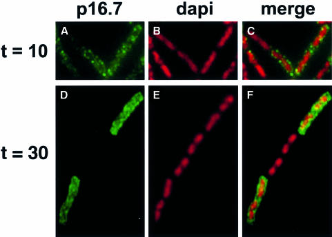
Fig. 5. Localization of protein p16.7 during the φ29 infection cycle. Aliquots of B.subtilis 110NA cells, harvested 10 min (A–C) or 30 min (D–F) after infection with sus14(1242) phage, were fixed and analysed by immunofluorescence using affinity-purified antibodies against p16.7. FITC immunofluorescence images (A and D) are shown in the left frames, corresponding DAPI images are shown in the middle frames (B and E) and combined images of both signals are shown in the right frames (C and F). Note that in the image shown at 30 min, the middle cell of the chain was probably not infected with φ29, so no p16.7 signal was detected.

An official website of the United States government
Here's how you know
Official websites use .gov
A
.gov website belongs to an official
government organization in the United States.
Secure .gov websites use HTTPS
A lock (
) or https:// means you've safely
connected to the .gov website. Share sensitive
information only on official, secure websites.
