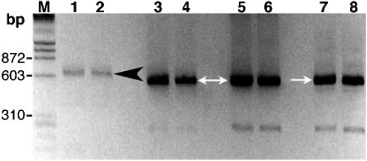
Fig. 2. PCR of trypanosomes in the patient’s blood.
Amplicons that included ITS1 of trypanosome-specific rDNA were generated with primers TRYP1S and TRYP1R and visualized on an agarose gel stained with ethidium bromide. The molecular size standard (M) was λ phage DNA digested with HindIII and ϕX DNA digested with HaeIII. Lanes: 1–2, blood sample from the Thai infant in duplicate assays; 3–4, T. evansi (Thailand); 5–6, T. evansi (Taiwan); 7–8, T. brucei rhodesiense (IL1501). The 520 and 623 bp amplicons are indicated by white arrows and a black arrowhead, respectively.

