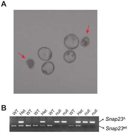
Figure 4. Snap23 Δ/Δ blastocysts die prior to uterine implantation.
(A) To evaluate the timing of embryonic lethality, embryos were collected from super-ovulated Snap23 Δ/wt females mated with Snap23 Δ/wt male mice by uterine flushing at E3.5. About 1/4 of the isolated blastocysts were morphologically abnormal and appeared to be degenerating; unlike sibling normal blastocysts they failed to develop any further during 24 hrs of culture (indicated by red arrows; see also Table 2). (B) Representative example of genotyping analysis revealing that abnormal blastocysts are homozygous for the Snap23 deleted allele (Snap23 Δ/Δ). Genomic DNA was isolated from individual blastocysts (shown in panel (A)) following 24 hr in culture, and genotyping was conducted using primers genoE2 SS, genoE2 AS, and genoE3 rev. PCR products for the Snap23 wt allele (266 bp) and for the Snap23 Δ allele (492 bp) are indicated.

