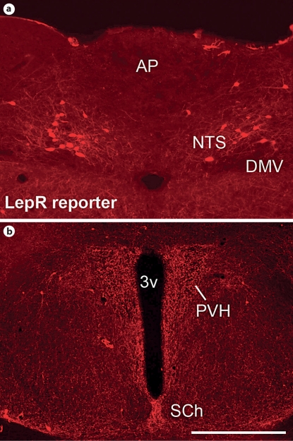
Fig. 2.
Distribution of neurons expressing LepR in the NTS and PVH. Neurons expressing LepR were visualized using LepR-IRES-Cre mice crossed with tdTomato reporter mice (B6.Cg-Gt(ROSA)26Sortm9(CAG-tdTomato)Hze/J, JAX® mice). a Fluorescent photomicrograph showing neurons expressing LepR in the NTS. Observe the close proximity with the AP. b Fluorescent photomicrograph showing fibers originating from LepR-positive neurons in the PVH. Observe the absence of cell bodies containing the reporter gene within the limits of the PVH. 3v = Third ventricle; DMV = dorsal motor nucleus of the vagus nerve; SCh = suprachiasmatic nucleus. Scale bar: a 200 μm; b 400 μm.

