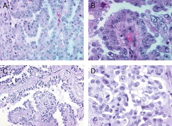
Figure 3.
Histopathology of the renal cancers. (A) The papillary structure of the renal cancer of patient FAM-1/IV-11 (H&E, magnification ×200). (B) Cells with inclusion-like nucleoli (patient FAM-1/III-9, H&E, magnification ×400). (C and D) The papillary structure and nuclear features of the lesion from patient FAM-2/II-2 [(C) H&E, magnification ×200; (D) H&E, magnification ×400].

