
Figure 1.
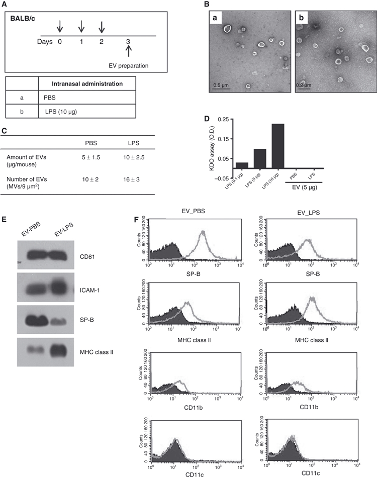
The production of extracellular vesicles (EVs) is enhanced by airway application of LPS, when compared with PBS application. (A) Protocol for EV preparation. (B) Transmission electron microscopy (TEM) images of EVs derived from the Bronchoalveolar lavage (BAL) fluids of PBS-treated (a) and LPS-treated (b) mice (N = 20 each group). (C) The amounts and numbers of EVs in the BAL fluids of PBS-treated and LPS-treated mice. The amounts of EVs were quantified on the basis of protein concentrations, as measured using the Bradford assay. The numbers of EVs were determined by counting the EVs in the TEM images. (D) The levels of LPS in the EVs isolated from the BAL fluids of LPS-treated and PBS-treated mice. (E) Western blotting assessment of the levels of host cell marker proteins, CD81, ICAM-1, surfactant protein B (SP-B), and MHC class II, in the EVs derived from LPS-treated and PBS-treated BALB/c mice. (F) FACS data of LPS- and PBS-induced EVs for the expression levels of the airway epithelial cell marker protein surfactant protein B (SP-B) and inflammatory cell marker proteins, MHC class II, CD11b, and CD11c. For (D–F): EV_PBS, EVs from PBS-treated mice; EV_LPS: EVs from LPS-treated mice.