Fig. 1.

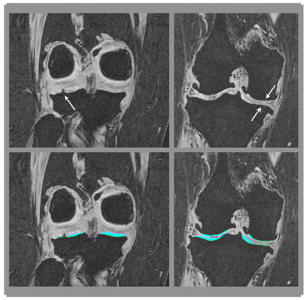
Different phenotypes of denuded areas of subchondral bone (dAB): internal osteophyte (left) and full thickness cartilage loss (right). The top row shows the un-segmented MR image, the bottom row the segmented MR image.

Different phenotypes of denuded areas of subchondral bone (dAB): internal osteophyte (left) and full thickness cartilage loss (right). The top row shows the un-segmented MR image, the bottom row the segmented MR image.