Fig. 2.
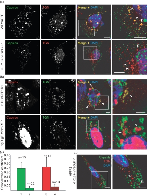
Association of capsids with the TGN. (a) HeLa cells were infected with 5 p.f.u. of vVP26GFP or vFRΔ37-VP26GFP per cell. At 15 h post-infection, the cells were fixed and stained with anti-TGN46 antibody and a GAR568 antibody (red). Capsids were visualized through direct GFP fluorescence (green). Bar, 10 μm. (b) HeLa cells were infected with 5 p.f.u. of vUL35RFP1D1 or vgD-gE-VP26RFP per cell. At 15 h post-infection, cells were fixed and labelled with anti-TGN46 antibody and a GARCy5 antibody (pseudo-coloured in green). Capsids were visualized through direct RFP fluorescence (red). In all cases, nuclei were stained with DAPI (blue). The boxed regions in the Merge+DAPI images are shown enlarged in the final panel and the positions of some TGN-derived vesicles are indicated by arrowheads. (c) Quantification of the amount of TGN signal that colocalizes with capsid signal for the four different viruses in HeLa cells (1, vVP26GFP; 2, vFRΔ37-VP26GFP; 3, vUL35RFP1D1; 4, vgD-gE-VP26RFP). The numbers of fields of view analysed are indicated above each bar. (d) HFFF2 cells were infected with 5 p.f.u. of vFRΔ37-VP26GFP per cell and labelled as above. A capsid aggregate is indicated by an arrow and a TGN vesicle by an arrowhead. Bars, 5 μm.

