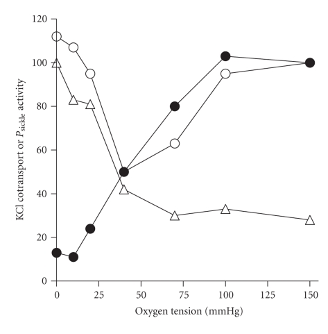
Figure 2.
Effect of oxygen tension on the activity of KCl cotransport (KCC) or P sickle in red blood cells (RBCs) from normal individuals or patients with sickle cell disease. The activity of each transport pathway is normalised—to the value in oxygenated cells (150 mmHg O2) for KCC activity and for that in deoxygenated RBCs (0 mmHg) in the case of P sickle—and given as a percentage. Solid circles give KCC activity in RBCs from normal HbAA individuals; open symbols give KCC activity (open circles) or P sickle activity (open triangles) in RBCs from sickle cell patients (HbSS homozygotes). In these experiments, total magnitude of KCC activity was about 10-fold greater in RBCs from HbSS individuals compared with HbAA ones. Note how the deoxygenation-induced KCC activity and activation of P sickle follow a similar dependence on O2 tension. Data taken from [67].

