Figure 2.
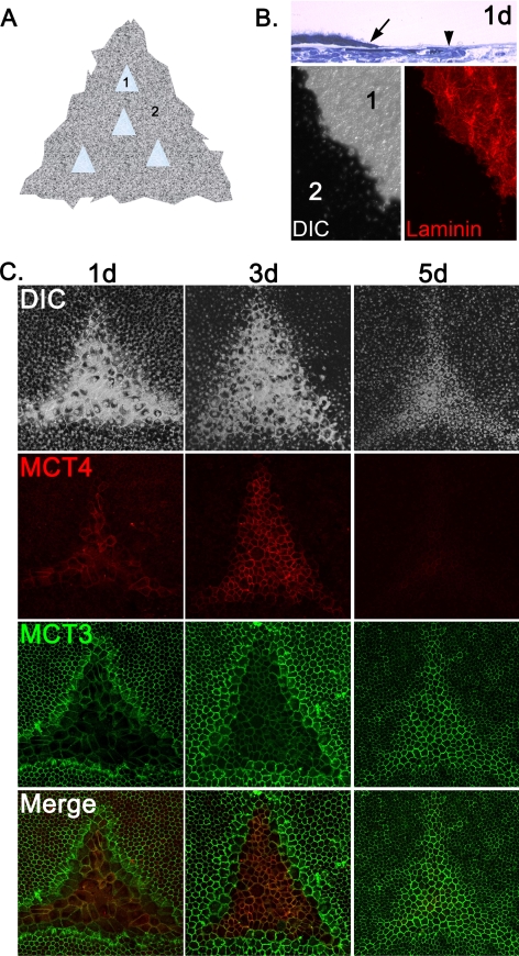
Wounding of RPE explants cause MCT isoform switching. (A) Schematic representation of wounding paradigm for chick RPE/choroid explant cultures. 1, wound region; 2, intact monolayer. (B, top) Histologic section showing the region of wounding of the chick RPE explant culture at 1 day (1d) after wounding. Arrow: wound edge. Arrowhead: intact basement membrane. Bottom: wounds were labeled with anti–laminin antibody to demonstrate that the basement membrane remained intact after wounding. 1 and 2 correspond to numbering in (A). Left: differential interference contrast. Right: laminin labeling. (C) At 1 day after wounding, a switch in MCT isoform expression was detected in the leading edge of the wound, where MCT4 was turned on and MCT3 was turned off. MCT4 expression was further enhanced in proliferating and migrating cells inside the wound at 3 days after wounding, whereas MCT3 expression remained low in this region. By 5 days after wounding, the wound region had redifferentiated and MCT3 was re-expressed.

