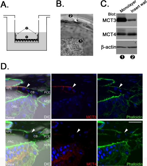
Figure 3.
The RPE requires continuous cell-cell contact to maintain MCT3 expression. (A) Schematic demonstrating migration of hfRPE cells in culture. 1, hfRPE monolayer; 2, hfRPE cells that migrated up the walls of the transwell insert. (B) Light micrograph demonstrating the movement of hfRPE up the wall of the transwell insert. Numbers correspond to those in (B). Arrow: area of migration up the wall of the insert. (C) Immunoblot analysis showing that MCT3 was expressed at high levels in lysates of hfRPE monolayers and at lower levels in lysates of hfRPE cells migrating up the sides of the transwell inserts. MCT4 expression was observed in each preparation. β-Actin, internal loading control. Numbers correspond to those in (B). (D) MCT3 expression was clearly limited to the RPE in the mouse eye. MCT4 was not expressed in this region. Arrow: border between the RPE and the ciliary epithelium. Blue: cells were stained to visualize nuclei. CB, ciliary body; PCE, pigmented ciliary epithelium; NCE, nonpigmented ciliary epithelium; PRC, photoreceptor cell; ONL, outer nuclear layer. Scale bar, 20 μm. Original magnification, 63× with a 2× zoom.

