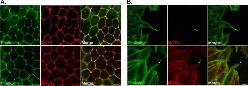
Figure 4.
Alteration in protein expression in wounded hfRPE cells. (A) Confocal immunofluorescence microscopy revealed that MCT3 (top) and MCT4 (bottom) co-localized with actin filaments at the lateral borders of hfRPE cells, indicative of basolateral polarity. (B) MCT3 expression was not detected at the leading edge of the wound (top, arrows). However, MCT4 expression (bottom) was observed both in cells at the leading edge of the wound (arrow) and at the lateral cell-cell borders (arrowhead). The cytoskeleton was visualized with phalloidin-488 labeling. Cells were fixed for immunofluorescence 48 hours after wounding. Scale bar, (A, B) 10 μm.

