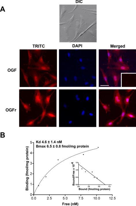
Figure 1.
The presence and distribution of OGF and OGFr in rabbit Tenon's capsule fibroblasts. (A) Photomicrographs of log-phase RTCFs visualized with differential interference (DIC) microscopy or immunocytochemistry of samples stained with antibodies (1:100) to [Met5]-enkephalin (OGF) or OGFr. Nuclei were visualized with DAPI. Rhodamine-conjugated IgG (1:1000) served as the secondary antibody (inset). Immunoreactivity was associated with the cytoplasm and the nucleus; staining was not observed in cell preparations incubated with secondary antibodies only (inset). Scale bar, 15 μm. (B) Representative saturation isotherm of specific binding of [3H]-[Met5]-enkephalin to nuclear homogenates of RTCFs. Mean ± SE binding affinity (Kd) and maximum binding capacity (Bmax) from four independent assays performed in duplicate. Representative Scatchard plot (inset) of specific binding of radiolabeled [Met5]-enkephalin to RTCF nuclear proteins revealed a one-site model of binding.

