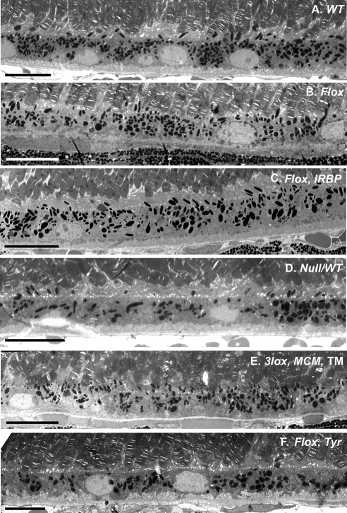
Figure 3.
Electron microscopy of the ChmWT (A), ChmFlox (B), ChmFlox, IRBP-Cre+ (C), Chm Null/WT (D), TM -induced Chm3lox, MerCreMer+ (E), ChmFlox, Tyr-Cre+ (F) at 5 months. In (A), (B), (C), and (E), the RPE is highly pigmented, and melanosomes are found in apical processes. In contrast, in (F) and some areas of (D), pigmentation is altered and melanosomes remain in the cell body. Scale bar, 10 μm.

