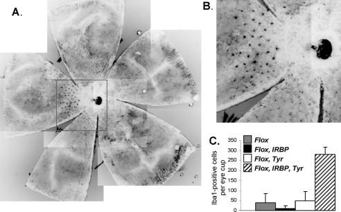
Figure 7.
(A) Retinal flat mounts from an 11-month-old ChmFlox IRBP-Cre+, Tyr-Cre+ animal were stained with anti–Iba1 primary and anti–rabbit Alexa-488 secondary antibody. (B) Enlargement of the boxed area in (A). (C) Quantitative data of the number of Iba1-positive cells per eye cup from 11- to 12-month-old ChmFlox (gray bar), ChmFlox, IRBP-Cre+ (closed bar), ChmFlox, Tyr-Cre+ (open bar), and ChmFlox, IRBP-Cre+, Tyr-Cre+ (hatched bar) animals.

