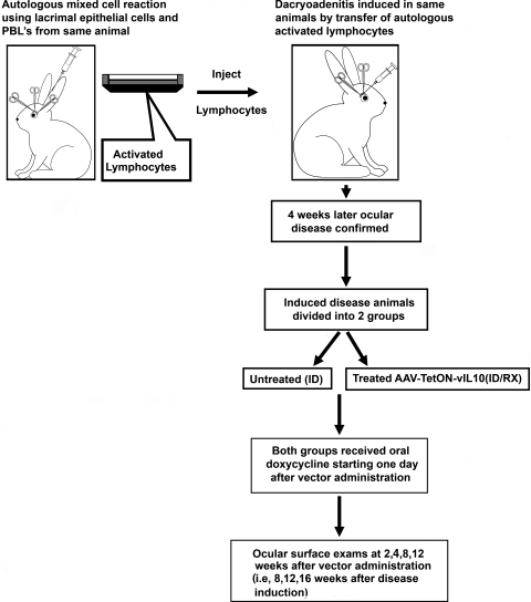
Figure 1.
Time sequence of experiments. Left: inferior lacrimal gland was removed from the rabbit, and autologous mixed-cell reaction was performed. Right: stimulated lymphocytes were injected into each donor rabbit's remaining lacrimal gland to induce dacryoadenitis. After 4 weeks, the first clinical assessments were performed. Once the disease was confirmed, the rabbits were randomly divided into two study groups. One group received AAV TetON-vIL-10 (ID/Rx), and the other group remained untreated (ID). Both groups received doxycycline in the drinking water (200 mg/kg body weight) from the next day of vector administration. The second clinical assessments were performed 2, 4, 8, and 12 weeks after vector administration (i.e., 6, 8, 12, and 16 weeks after disease induction).

