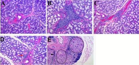
Figure 4.
Histopathology features of treated and untreated animals. (A) Normal LG showed occasional small lymphocytic aggregates. (B) After 16 weeks, dacryoadenitis induction resulted in substantial lymphocytic infiltration in the LG, primarily periductal and perivenular. (C) Sections from ID/Rx 12 weeks after the treatment period revealed small to medium foci around ducts and venules that were considerably smaller and less frequent than in the ID group. (D) LG of rabbit 227 from the ID/Rx group was largely normal in appearance and contained small lymphocytic foci (red arrow). (E) One area of the rabbit 227 gland was dramatically altered, presenting with clusters of duct-like structures (circles) and evidence of streaming lymphocytes (black arrowhead) suggesting proliferation.

