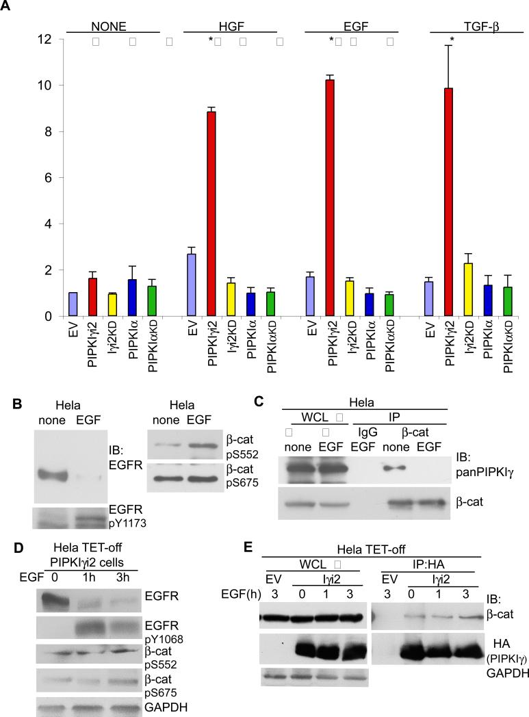
Figure 5. PIPKIγ activates β-catenin upon growth factor stimulation.
(A) β-catenin transcriptional activity was measured in Hela cells transiently transfected with empty vector (EV) or the HA-tagged PIPKI shown. 24 h after transfection, cells were serum starved and then left untreated (none) or stimulated with 1nM EGF, 50 ng/ml HGF or 2ng/ml TGF-β for 24 h where indicated followed by cell lysis to measure β-catenin activity (n≥3, error bars=std dev, * represents p ≤ 0.01). (B) Hela cells were serum starved and then left untreated (none) or stimulated with 1nM EGF for 2 h where indicated. Whole cells lysates (WCL) were prepared and probed with the antibodies shown. (C) Cells were treated as in (B). Following cell lysis, equal protein concentrations from none and EGF treated whole cell lysates were incubated with anti-β-catenin antibody. Immunoprecipitated β-catenin and co-IPed PIPKIγ were detected with specific antibodies. (D) Hela TET-off PIPKIγ_i2 cells were maintained in medium without doxycycline to induce PIPKIγ_i2 expression. Cells were serum starved and stimulated with 1nM EGF for 0, 1 or 3 h where indicated. Whole cells lysates (WCL) were prepared and probed with the antibodies shown. (E) Cells were treated as in (D). Following cell lysis, whole cell lysates of equal protein concentrations were incubated with anti-HA antibody. Immunoprecipitated HA-PIPKIγ_i2 and co-IP'd β-catenin were detected with specific antibodies. Hela TET-off cells transfected with the empty vector were used as a control during the IP.

