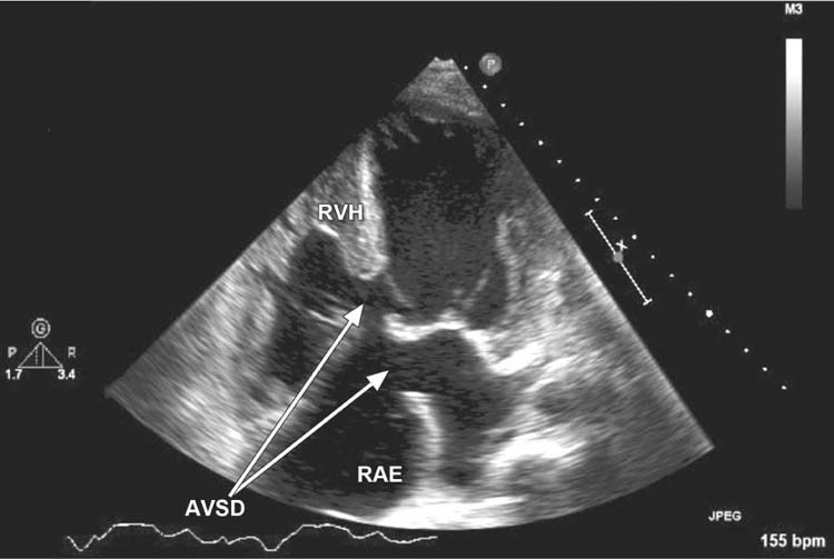
Fig. 1 Transthoracic echocardiogram (apical 4-chamber view) shows complete atrioventricular canal defect with secondary pulmonary hypertension. Note the large atrioventricular septal defect (AVSD), right ventricular hypertrophy (RVH), and right atrial enlargement (RAE).
