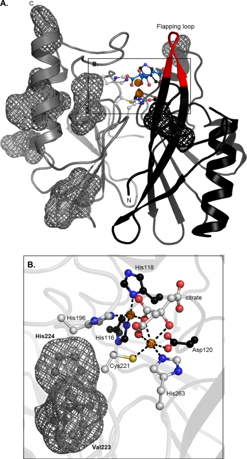
FIG. 4.
Structure of VIM-4. (A) Three-dimensional structure of VIM-4 enzyme. Both Zn2+ ions are represented as orange spheres, Zn2+ ligands (gray or black sticks) are shown, and the locations of the VIM-4/VIM-2 mutations are shown as meshes. (B) Active site of the dizinc structure. Citrate and ligands are represented as sticks. The figures were generated with the PyMOL program.

