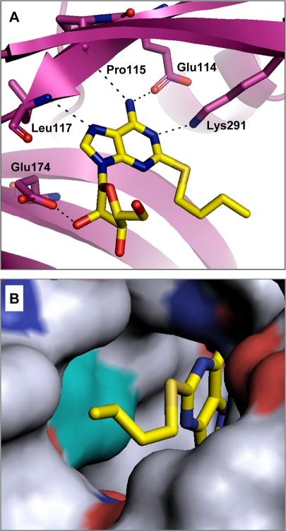
FIG. 3.
(A) X-ray cocrystal structure of compound 1 bound to the AMP-binding site of H. influenzae LigA. Predicted hydrogen bonds between the inhibitor and the enzyme are shown as dashed lines. Enzyme residues proposed to make key interactions with the compound are labeled. (B) Surface representation of the hydrophobic tunnel of the binding pocket containing compound 1. The surface of L82 (corresponding to L75 in S. pneumoniae LigA) is shown in cyan.

