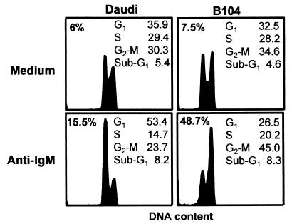
Figure 1.
The effect of anti-IgM on the viability and cell cycle distribution of B104 and Daudi cells. Cells (5 × 10⁵) were incubated for 24 h in the presence or absence of 20 μg/ml anti-IgM, stained with PI before and after permeabilization, and analyzed by flow cytometry. The percentage of dead cells was detected by PI exclusion (upper left corners), and the proportion of cells in each phase of the cell cycle and in sub-G1 phase was calculated according to the DNA content. This is a representative experiment of three experiments performed.

