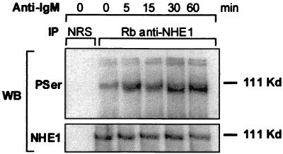
Figure 4.
The levels of NHE1 phosphorylation in anti-IgM-treated Daudi cells. Cells were incubated with 20 μg/ml anti-IgM for various times as indicated. NHE1 immunoprecipitates and control precipitates were electrophoresed on a 7.5% SDS/PAGE, transferred to poly(vinylidene difluoride), and immunoblotted as described in Fig. 3. This is a representative experiment of three experiments performed.

