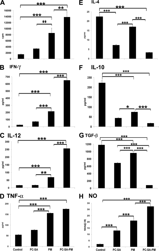
FIG. 4.
LAg-specific lymphoproliferation, cytokine, and NO levels in differently treated infected mice. Spleen cells of indicated treated animals were isolated 4 weeks posttreatment, plated aseptically (2 × 10⁵ cells/well), and stimulated with LAg at 2 μg/ml for 48 h. (A) LAg-specific in vitro proliferation of spleen cells of differently treated animals was determined. IFN-γ (B), IL-12 (C), TNF-α (D), IL-4 (E), IL-10 (F), TGF-β (G), and NO (H) in spleen cell culture supernatants of indicated treatment groups were determined by ELISA (for panels B to G) and the Greiss assay method (for panel H). Data represent the mean ± SE for five animals per group. Data were tested by ANOVA. Differences between means were assessed for statistical significance by Tukey's test (**, P < 0.01; ***, P < 0.001).

