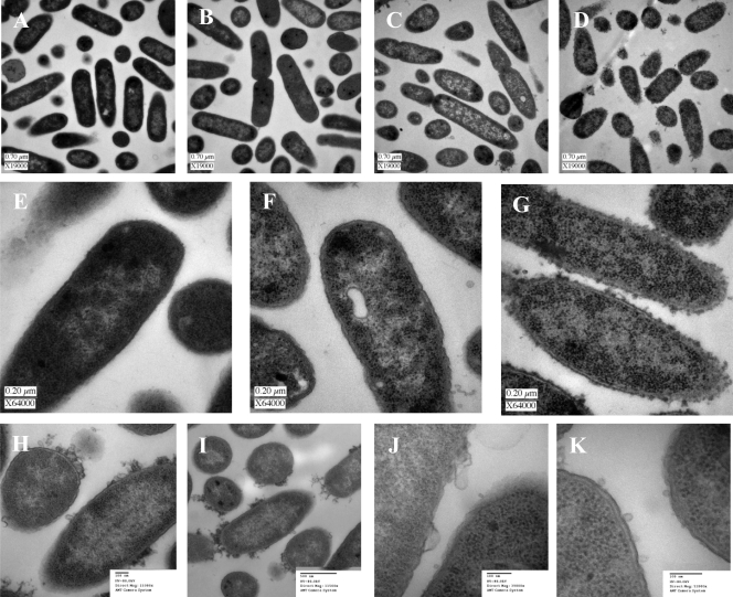
FIG. 3.
Transmission electron microscopy findings. Logarithmically growing E. coli cells were cultured in MOPS minimal medium in the presence or absence of various concentrations of Bac8c. Samples were taken after 30 min, and cells were fixed in glutaraldehyde immediately and then processed for TEM. (A) Control; (B) 3 μg/ml (IC50); (C) 6 μg/ml (MBC); (D) 30 μg/ml (5× the MBC). (E to G) At a higher magnification, the control (E), cells exposed to 6 μg/ml Bac8c (F), and cells exposed to 30 μg/ml Bac8c (G) are shown. (H to K) Alternate images of cells at the MBC (6 μg/ml), focused on the membrane effects of Bac8c.

