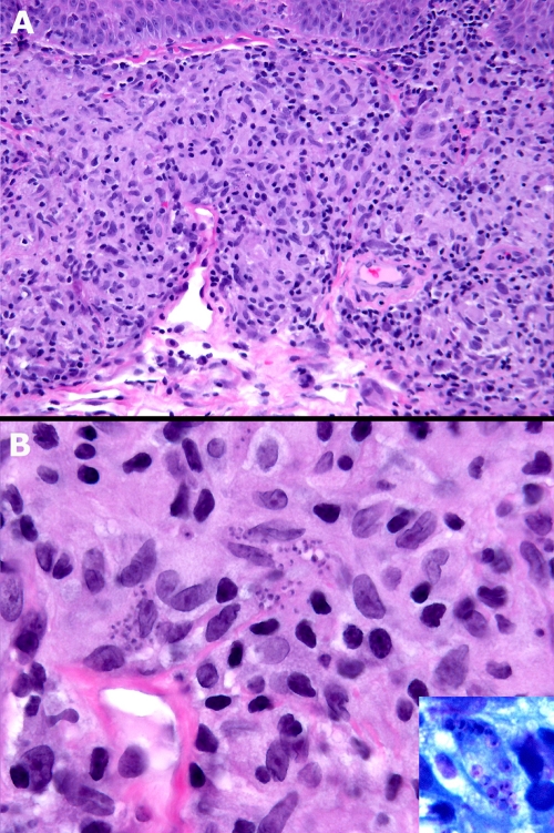
FIG. 1.
(A) Diffuse mixed granulomatous dermal infiltrate, mainly lymphoid-histiocytic (hematoxylin and eosin stain; magnification, ×20). (B) Clusters of basophilic microorganisms (amastigotes) within the histiocytes (hematoxilin and eosin stain; magnification, ×60). Inset, amastigotes within a vacuolated histiocyte. The nuclei of L. infantum are clearly visible (Giemsa stain; magnification, ×100).

