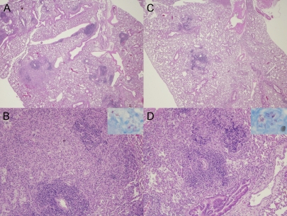
FIG. 1.
Lung histopathology in the murine model of chronic TB infection following pyrazinamide therapy. (A and B) Day 0 of treatment (28 days after aerosol infection). Magnifications: A, ×2; B, ×10. Interstitial fibrosis and marked pulmonary edema in the alveolar spaces. Cellular lesions are seen comprising mainly lymphocytes with few histiocytes and plasma cells (H&E stain). (Inset in panel B) AFB are detected within cells. (C and D) Day 28 after treatment with pyrazinamide at 150 mg/kg. Magnifications: C, ×2; D, ×10. Interstitial lymphocytic infiltrates surrounded by areas of pulmonary edema (H&E stain) can be seen. (Inset in panel D) AFB are confined to the intracellular compartment. Well-formed granulomas were not seen at either time point in any group.

