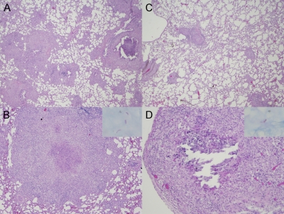
FIG. 2.
Lung histopathology in the guinea pig model of chronic TB infection after pyrazinamide therapy. (A and B) Day 0 of treatment (28 days after aerosol infection). Magnifications: A, ×2; B, ×10. Granulomas with punctuate necrosis and large areas of epithelioid histiocytes (H&E stain) are visible. (Inset in panel B) AFB are detected primarily within histiocytes. (C and D) Day 28 after treatment with pyrazinamide at 300 mg/kg. In panel C a decrease in the number and size of granulomas, which are localized more peripherally in the lungs (H&E stain, ×2 magnification), can be seen. Panel D shows a caseating granuloma with partially missing necrotic center, encircled by epithelioid histiocytes and lymphocytes (H&E stain, ×10 magnification). In the inset, AFB are extracellular and confined to areas of necrosis.

