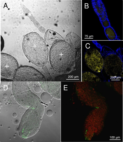
FIG. 7.
Visualization by CSLM of the gut of nymphs and female gonads of H. obsoletus. (A) Interferential contrast microscopy image of a female gonad. (B and C) DAPI (4′,6-diamidino-2-phenylindole) staining and FISH with the Cy5-labeled W1 probe specific for Wolbachia (yellow). Insect cell nuclei stained with DAPI are blue. Magnifications of an immature ovariole (asterisk) (B) and of a mature egg (plus) (C) are shown. (D) Interferential contrast microscopy image of a nymphal gut overlapped with an FITC-labeled W2 probe specific for Wolbachia (green). (E) The same image after propidium iodide staining and FISH using the FITC-labeled probe W2 specific for Wolbachia (green).

