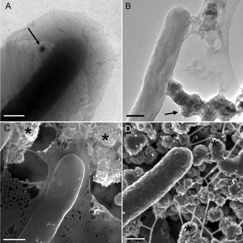
FIG. 2.
Comparison of bacteria and EPS in the hydrated and dehydrated states. Resting-cell suspensions of S. oneidensis MR-1 were incubated anaerobically for 24 h with lactate and MnO2, and representative assay aliquots were prepared for concurrent cryo-EM and RT EM imaging. (A) Cryo-TEM micrograph of cell illustrating EPS in the hydrated state. The slight contrast of the EPS is likely due to bound Mn(II) resulting from dissimilatory reduction of Mn(IV). The arrow in panel A shows the MnO2 crystalline material. (B) The whole-mount RT TEM preparation shows a cell with the collapsed EPS covering the MnO2 material (arrow). (C) Representative cryo-SEM image of rapidly frozen material after partial sublimation displays partially hydrated cells associated with EPS that covers the MnO2 material (asterisks). (D) A fixed, dehydrated, and CPD-prepared sample characteristically shows excellent 3D cellular preservation but completely altered EPS that forms fiber-like structures colocalized with MnO2 material (asterisks). Scale bars: 500 nm.

