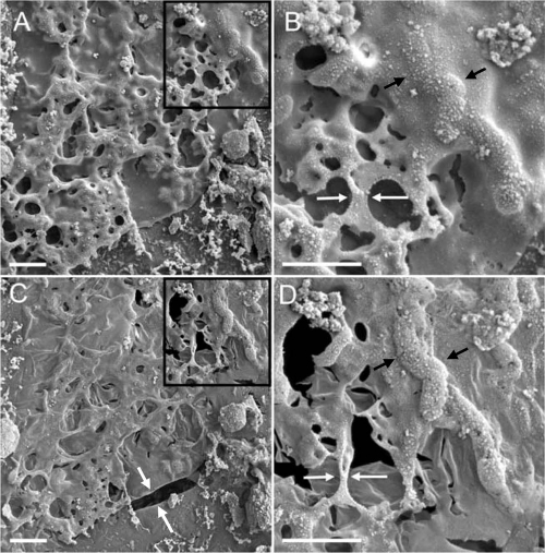
FIG. 4.
Process of gradual moisture loss from frozen-hydrated natural biofilm as captured by cryo-SEM. (A and B) Initial image obtained at −180°C in the fully hydrated state after the biofilm was plunge-frozen. The black arrow in panel B points to a contour of bacteria embedded in EPS material that remains partially hydrated. (C and D) The same area of biofilm after sequential warming to −150°C. Notice the substantial collapse in the z direction (thickness), and beginning of the shrinkage in the x and y dimensions, resulting in cracks (white arrows in panel C). White arrows in panels B and D highlight the structural alteration of EPS, with the formation of a filamentous structure. The cellular features in panel D (black arrows) are more pronounced after sublimating a thin layer of the water from the surface. Black boxes in panels A and C illustrate the areas shown in higher magnification in panels B and D, respectively. Scale bars: 10 μm in panels A and C and 2 μm in panels B and D.

