FIG. 5.
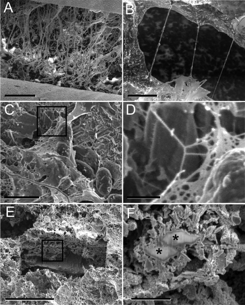
Observation of biofilm features by cryo-SEM and RT SEM. (A and B) EPS transformation during sublimation induced by progressive warming in the cryo-SEM. The biofilm shows substantial structural damage due to material collapse and contraction induced by warming to −60°C. (A) Distortion of biofilm surface features with extensive cracks (>200 μm) developed over time in the initially cohesive material. (B) Formation of filamentous structures could be observed in situ during the surrounding material shrinkage, while the partially hydrated viscoeleastic EPS material was stretched between apparent anchored points. (C and D) Morphologically similar filamentous structures formed during sublimation were also observed in samples prepared by CPD and viewed at RT in the SEM. (E) Cryo-FIB milling of an approximately 20-μm depth below the biofilm surface revealed superior structure preservation. (F) Cryo-FIB-prepared area with mineral-laden layers of EPS and cells in various degrees of mineralization (asterisks), with no signs of EPS collapse and stretching. Black boxes in panels C and E illustrate the areas shown in higher magnification in panels D and F, respectively. Scale bars: 200 μm (A), 5 μm (B), 2 μm (C), 500 nm (D), 30 μm (E), and 2 μm (F).

