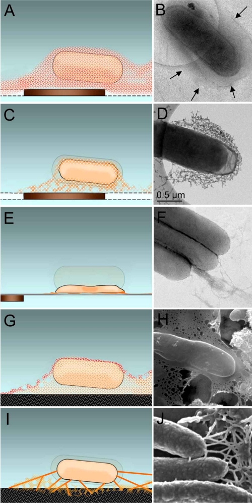
FIG. 6.
Model and correlated EM images of cells and associated EPS structural alteration resulting from different methods of sample processing. (A and B) Cryo-TEM of frozen-hydrated cells vitrified in amorphous ice imaged at −178°C. Arrows indicate the outline of the EPS. (C and D) RT TEM of initially vitrified cells that have been gently dried in low vacuum of the DPS. (E and F) RT TEM of air-dried cells stained with Nano-W (Nanoprobes, Yaphank, NY) prior to imaging. (G and H) Cryo-SEM of cells in a thin layer of partially sublimated amorphous ice. (I and J) RT SEM of cells that were fixed, dehydrated, and prepared by CPD. Panels C, E, and I also show hydrated cell outline for comparison of cell shrinkage.

