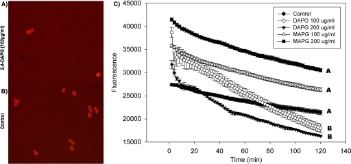
FIG. 3.
Interrupted cell homeostasis in wild-type strain BY4741 by 2,4-DAPG. Cells treated with 2,4-DAPG or MAPG at 100 or 200 μg ml−1 were exposed to acridine orange for 2 h at 30°C. (A) Cells treated with 100 μg ml−1 2,4-DAPG; (B) control, no 2,4-DAPG added; (C) acridine orange fluorescence intensities. Each point of each treatment is the mean of results from four replicates. Fluorescence was measured every 2 min with a SAFIRE microplate reader (excitation at 502 nm, emission at 525 nm). Slopes of curves were calculated using the linear regression function in SigmaPlot 8.0. Lines with the same letters are not significantly different according to Tukey's HSD test (P = 0.01).

