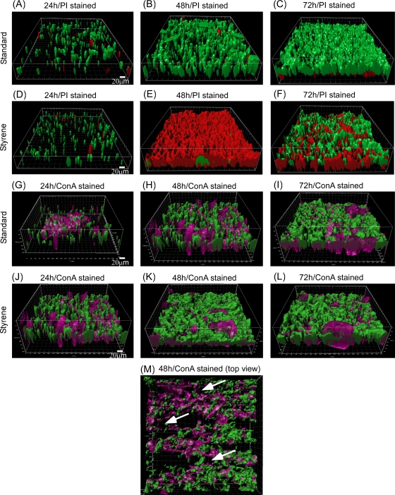
FIG. 1.
Confocal micrographs showing biofilm development stages under standard growth conditions (no solvents) and in the styrene environment. (A to C) gfp-expressing intact and PI-stained biofilms under normal growth conditions after 24 (A), 48 (B), and 72 (C). (D to F) gfp-expressing intact and PI-stained biofilms in the styrene environment after 24 (D), 48 (E), and 72 h (F). (G to I) ConA-stained biofilm under normal growth conditions after 24 (G), 48 (H), and 72 h (I). (J to L) ConA-stained biofilm in the styrene environment after 24 (J), 48 (K), and 72 h (L). (M) Top view of 48-h-old ConA-stained biofilm with water channels as indicated by arrows. Green color represents the intact gfp-expressing cells, red color represents PI-stained dead or permeabilized cells, and violet color (ConA) represents polysaccharides in the EPS matrix. Representative IMARIS-treated and 3D-reconstructed images from three independent experiments are shown. Scale bar, 20 μm.

