FIG. 4.
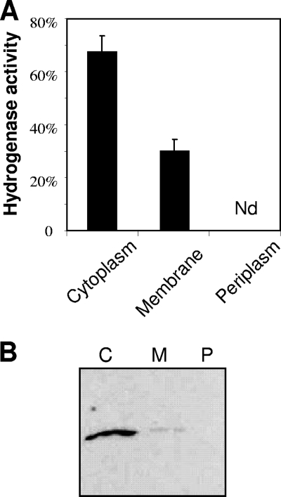
Determination of A. macleodii HynSL localization. (A) Detection of A. macleodii HynSL activity in the cytoplasm, membrane, and periplasm fractions. Bars depict relative activity with respect to the total activity prior to cellular fractioning (100% activity). The data displayed represent average values ± the SD of three independent experiments. Nd, none detected. (B) Immunodetection of A. macleodii HynSL in the cytoplasm, membrane, and periplasm fractions. Three cellular fractions prepared from equal amounts of cells were separated on a native PAGE, and the protein blot was probed with anti-TrHynL antibodies. Lanes: C, the cytoplasmic fraction; P, the periplasmic fraction; M, the membrane fraction. A total of 40 μg of total protein was loaded into each lane for Western blotting.

