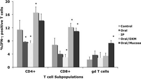
FIG. 2.
Percentages of IFN-γ+ T cells among PBMCs isolated from control noninfected calves and calves infected with Mycobacterium avium subsp. paratuberculosis by the following methods: oral, intraperitoneal, oral with dexamethasone administration, and oral with mucosal scrapings from a cow with clinical disease. Percentages of IFN-γ+ T cells were determined at 12 months for CD4, CD8, and γδ T-cell subpopulations stimulated with a whole-cell sonicate of Mycobacterium avium subsp. paratuberculosis (MPS). Data are expressed as means ± SEM. Significant differences between control and infection groups within the 12-month time point are represented by asterisks (*, P < 0.05).

