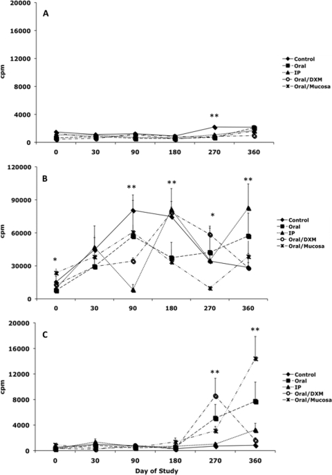
FIG. 7.
Lymphocyte proliferation responses (cpm) by PBMCs isolated from control noninfected calves (⧫) and calves infected with Mycobacterium avium subsp. paratuberculosis by the following methods: oral (▪), intraperitoneal (▴), oral with dexamethasone administration (○), and oral with mucosal scrapings from a cow with clinical disease (✳). Lymphocyte proliferation responses during the 12-month study were measured in nonstimulated PBMCs (A), PBMCs stimulated with ConA (B), and PBMCs stimulated with a whole-cell sonicate of Mycobacterium avium subsp. paratuberculosis (MPS) (C). Data are expressed as means ± SEM. Significant differences between control and infection groups within given time points are represented by asterisks (**, P < 0.01; *, P < 0.05).

