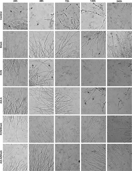
Fig. 1.
Images of P. brasiliensis during M-to-Y differentiation in the presence of mitochondrial respiratory chain inhibitors. M-to-Y differentiation was induced as described in Materials and Methods. Mitochondrial respiratory chain inhibitors were added just before the temperature was shifted from 26°C to 35.5°C. Their concentrations per addition, even when combined, were as follows: for SHAM, 2 mM; for KCN, 1 mM; and for antimycin A, 1.8 μM. In some of the experiments, drugs were added once more after 24 or 72 h, but there were no noticeable differences between the results obtained with single additions and those obtained with multiple additions (data not shown). Images were selected from a single experiment that was representative of at least three independent experiments. Images were captured using a 40× objective lens, resulting in a final magnification of ×400, with an Olympus BX51 model U-LH100-3 microscope that was coupled to an Olympus model C-5060 wide-zoom digital camera. Black arrows indicate chlamydospores, which are characteristic structures during M-to-Y differentiation.

