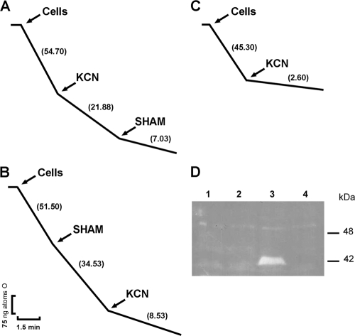
Fig. 8.
Cyanide-resistant respiration in Pbaox-expressing E. coli. (A and B) E. coli/pET28-Pbaox-induced cells. (B) E. coli/pET28. Cells were incubated at 30°C in 1.8 ml of a respiration medium. At the indicated time points, KCN (1 mM) and SHAM (2 mM) were added. The rate of oxygen uptake was expressed as ng atoms of oxygen/min. All the assays were performed with 0.2 mg/ml of proteins. The depicted graphs are representative of at least three independent assays. (D) Reverse image of the immunodetection of PbAOX in Pbaox-expressing E. coli. Fifty micrograms of protein was loaded per lane. Samples were prepared, as described in Materials and Methods, from induced E. coli cells that had been transformed with the empty vector (lane 1), uninduced E. coli cells that had been transformed with the empty vector (lane 2), induced E. coli cells that had been transformed with pET28-Pbaox (lane 3), and uninduced E. coli cells that had been transformed with pET28-Pbaox (lane 4). SDS-PAGE and Western blot techniques were performed as described in Materials and Methods. His-tagged PbAOX was detected using a monoclonal antibody that was raised against the His tag. The expected molecular mass for the final protein is about 42 kDa.

