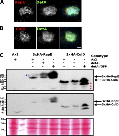
Fig. 8.
Putative DetA interaction partners. (A) Immunofluorescence on fixed detA−/GFP-DetA cells supertransformed with a 3×HA-RepE construct; (B) Immunofluorescence on fixed detA−/GFP-DetA cells supertransformed with a 3×HA-CulD expression construct; (C) Western blot showing Ax2, detA− or detA−/GFP-DetA cells supertransformed with the expression construct 3×HA-RepE or 3×HA-CulD. The middle panel is a shorter exposure of the top panel, and the lower panel shows the Ponceau-S-stained membrane to confirm equal loading. The red asterisks indicate putative degradation products of 3×HA-CulD, and the blue asterisk indicates a species of 3×HA-RepE with a higher than predicted Mr, possibly due to ubiquitination or another posttranslational modification. Scale bar, 1 μm.

