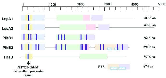
Figure 5.
Analysis of PfhB domains. Homology comparisons among PfhB1, PfhB2, from Pm, LspA1, LspA2, from H. ducreyi, FhaB from B. pertussis, and P76 from Haemophilus somnus are presented. Homologous domains are represented with the same colored boxes and the direct repeats in p76 and PfhA2 are patterned in blue. The N(P/Q)NG(I/M) extracellular processing motif is indicated and the integrin-binding protein motifs are shown as dark purple lines.

