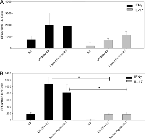
FIG. 1.
C. muridarum antigen-specific Th1 and Th17 cells are detected in similar numbers in the iliac nodes of C57BL/6 mice on day 7 postinfection, but Th1 cells predominate on day 20. The numbers of antigen-specific IFN-γ- and IL-17-producing CD4+ T cells in iliac nodes (ILN) of C57BL/6 mice were quantified by ELISPOT on day 7 (A) and day 20 (B) postinfection. Significantly increased numbers of IFN-γ-producing CD4+ T cells and IL-17-producing CD4+ T cells were noted on both day 7 (A) and day 20 (B) postinfection after stimulation with UV-EBs or C. muridarum pooled peptide compared to stimulation with IL-2 alone (P < 0.05 by one-way ANOVA). On day 7 postinfection, the numbers of UV-EB- and pooled-peptide-specific Th1 (IFN-γ-producing) and Th17 (IL-17-producing) cells were not significantly different. By day 20, the numbers of UV-EB- and pooled-peptide-specific Th1 (IFN-γ-producing) cells were significantly greater than those of Th17 (IL-17-producing) cells (*, P < 0.05 by one-way ANOVA). The data are representative of two individual experiments in which the cells from three groups of three mice each were pooled and analyzed. The error bars indicate SD. SFCs, spot-forming cells.

