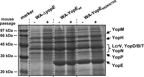
FIG. 2.
Yop secretion profiles of the investigated Yersinia strains. The Coomassie blue-stained SDS-polyacrylamide gel shows proteins released by Yersinia upon Ca2+ removal into the bacterial growth medium. The spectra of Yop release were analyzed for the YopE-negative mutant WA-ΔyopE and the mutant strain recomplemented with either wild-type YopE (WA-YopEwt) or K62- and K75-mutagenized YopE (WA-YopEK62R/K75Q) before and after the passage through mice. For mouse passage, the bacteria were recovered from the spleens of orogastrically infected mice at day 3 postinfection. Molecular size marker proteins are shown in the first lane.

