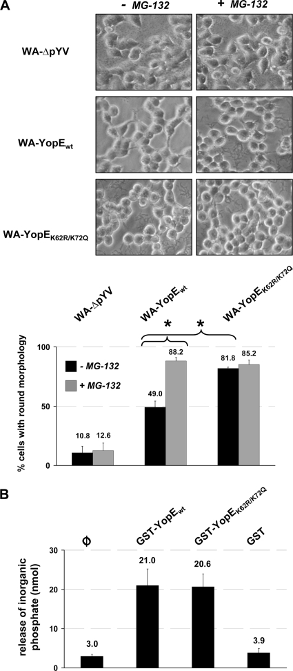
FIG. 4.
Wild-type YopE O8 triggers diminished, proteasome inhibitor-sensitive cytotoxic alterations of infected cells. (A) Differential sensitivity of YopE-conferred cytotoxicity to proteasome inhibition. HEK293 cells were left untreated or treated with the proteasome inhibitor MG-132 prior to infection with virulence plasmid-cured yersiniae (WA-ΔpYV) or yersiniae producing either wild-type (WA-YopEwt) or K62- and K75-mutagenized (WA-YopEK62R/K75Q) YopE O8. Ninety minutes after onset of infection, the yersiniae were killed by addition of gentamicin. The cells were fixed, and cellular morphologies were microscopically analyzed after a total incubation period of 4.5 h. The numbers of cells with a completely rounded phenotype were quantified from three separate experiments, and mean percentages of rounded versus total numbers of cells ± SD are indicated. Differences in cell rounding were statistically significant for WA-YopEwt versus WA-YopEK62R/K75Q in the absence of MG-132 and for WA-YopEwt with versus without MG-132 (P < 0.005). (B) Comparable in vitro GAP activities of YopEwt and YopEK62R/K75Q. Recombinant, purified GST-fused YopEwt and YopEK62R/K75Q were subjected to in vitro GAP assay using recombinant RhoG. GTP hydrolysis and release of free phosphate were measured at 650 nm after coincubation of RhoG and the YopE protein species for 20 min at 37°C. GST protein was used as negative control. φ, background phosphate levels in the absence of GST or GST-YopE. Specific GAP activity from two independent, representative experiments was determined with a phosphate standard and is indicated as mean nmol ± SD. The increase in GAP activity triggered by GST-YopEwt and GST-YopEK62R/K75Q compared to background levels (φ) was statistically significant (P < 0.03).

