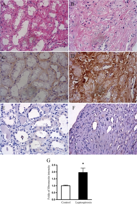
FIG. 2.
Increased deposition of fibronectin immunostaining in the tubulointerstitium of patients with leptospirosis. The typical renal histology of H&E staining in five leptospirosis patients is shown in panel B. Tubulointerstitial fibronectin deposition in these patients was assessed by immunostaining for fibronectin, as shown in panel D. Normal tissues cut from residual kidney specimens obtained from five nephrectomized patients with renal cell carcinoma were used as the control and stained for H&E staining (A) or fibronectin immunostaining (C). (E and F) A negative control from normal tissues (E) and diseased tissues (F) for fibronectin immunostaining was performed using only the secondary antibody with omission of the primary antibody (anti-fibronectin). (G) The integrated image intensity of fibronectin in eight consecutively selected nonoverlapping and area-fixed fields at a ×400 magnification in each kidney specimen was calculated as described in Materials and Methods. The results are presented as the relative fold immunostaining intensity over that of the control ± the SE (*, P < 0.05, leptospirosis patients versus control patients).

