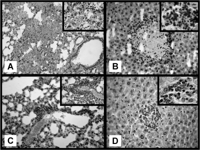
FIG. 7.
Histological examination in AP mice (hematoxylin-eosin staining). (A) Day 1 after AP, intra-alveolar lung hemorrhage. Magnification, ×250. (Inset) Destroyed alveolar septa. Magnification, ×400. (B) Nine days after AP, focus of hepatic necrosis surrounded by abundant leukocyte infiltration. Magnification, ×250. (Inset) Mixed inflammatory infiltrate with neutrophils and lymphocytes. Magnification, ×1,000. (C) Twenty days after AP, lung with focus of perivascular inflammatory infiltrate. Magnification, ×250. (Inset) A predominantly lymphocytic infiltrate. Magnification, ×400. (D) Twenty days after AP, focus of regenerated hepatocytes with chronic inflammatory infiltrate. Magnification, ×250). (Inset) Predominantly lymphocytic infiltrate. Magnification, ×1,000.

